
-
Sale!
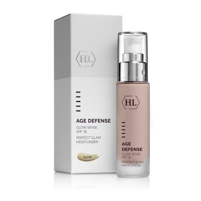
AGE DEFENSE GLOW SENSE SPF 15 50ml
Original price was: 58,66 €.44,00 €Current price is: 44,00 €. Add to cart -
Sale!
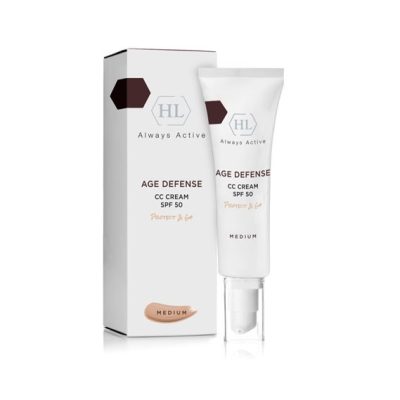
AGE DEFENSE CC CREAM SPF 50 MEDIUM 50ml protect & go ***NEW *** Limited Edition
Original price was: 51,36 €.38,52 €Current price is: 38,52 €. Add to cart -
Sale!
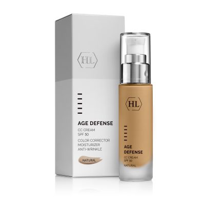
AGE DEFENSE CC CREAM SPF 50 NATURAL 50ml
Original price was: 59,43 €.44,57 €Current price is: 44,57 €. Add to cart -
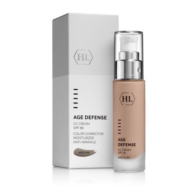
AGE DEFENSE CC CREAM SPF 50 MEDIUM 50ml
59,43 € Add to cart
Structure and function of the epidermis
The epidermis layer is the external layer of the skin, which protects the inner tissues from harmful external effects, such as air pollution, smoke, ultraviolet rays and dryness, all which cause an accumulation of free radicals in the body. The epidermis is composed of three main types of cells: keratinocytes, melanocytes, and Langerhans’ cells.
The keratinocyte cells, which constitute the majority of the cells, are arranged in layers. The upper layer is called the corneum layer, the intermediate layer is made up of elongated squamous cells, while the bottom layer is called the basal cell layer. Because the upper layer of the skin is constantly exposed to harmful external effects, the skin has a special mechanism to replace the upper skin cells every 28 days or more. Cells in the basal layer which is at the bottom of the epidermis move upwards. In the process of moving they die and form the corneum layer, which is in effect made up of dead cells that adhere to one another strongly with a type of biological adhesive, serving to prevent the penetration of harmful bodies such as bacteria and viruses.
At the bottom of the epidermis are melanoctye cells. These cells form melanin, a chemical substance that gives the skin its color. The number of melanoctyes is the same among light and dark-skinned people, but in dark skins, the melanin is formed continuously and is spread through all layers of the epidermis, while in light skin, it is formed only after continued exposure to the sun, and then too only in relatively small quantities, and is distributed only in the lower epidermis layers. In addition, the melanin that is formed in dark-skinned people is more effective in blocking the sun’s rays than that formed in light-skinned people.
The Langerhans’ cells are found in the upper epidermis layers and constitute an important part of the protective system of the skin and in fact of the whole body. These cells belong to the body’s immune system, and their role is to identify foreign substances that penetrate the skin, such as bacteria, viruses, or carcinogenic cells. In these cases, the Langerhans’ cells secrete special substances that summon to the “attacked’ region white blood cells – lymphocytes. The lymphocytes, which are the “soldiers” of the immune system, create an inflammatory reaction, which aims to destroy the foreign bodies.
Structure and function of the dermis
The dermis layer is responsible for the supply of support and food material to the epidermis. This layer is primarily composed of collagen and elastin fibers. With age, these fibers begin to change, and they give the skin an old and wrinkled look. The collagen is a type of protein built as thick and interwoven fibers, facing in different directions. In this way, they keep the skin strong and allow it to be stretched without tearing.
The elastin fibers are what gives the skin its elasticity. These fibers are relatively thin, and they behave like rubber, allowing the skin to return to its original form.
The dermis contains blood vessels that nourish the skin and provide it with food and oxygen. In addition, it contains the sebaceous glands, sweat glands, hair follicles, and sense receptors.
The sebaceous glands secrete an oily substance that coats the skin and keeps it moist. These glands are generally concentrated in the skin of the face, scalp, back, and chest. While present in the body from birth, the glands only become active during puberty.
The function of the sweat glands is to secrete sweat, in order to regulate the body temperature and cool it. The hair follicles are like small pouches, with a single hair growing in each. The sense receptors are responsible for the various senses of our skin – heat, cold, pain, touch, and pressure and thanks to them the skin is a vital organ in receiving information from the environment.
Sun protection
Recent studies have proven that exposure to the sun’s rays harms the functioning of the Langerhans’ cells found in the epidermis, which are vital for the immune system. This damage constitutes one of the main causes in the development of tumors on the skin related to the sun, among them cancerous tumors. This is the reason that today cosmetic companies are meticulous about including sun filters in their treatment products. HL’s SUNBRELLA and AGE DEFENSE products include sunscreens that provide comprehensive protection against UVA and UVB rays .